Mitral valve stenosis classification and possible complications of the disease
Content
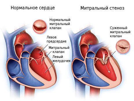
Mitral stenosis is a physiological disorder of the cardiac organ, characterized by a pathological narrowing of the area of the left atrioventricular orifice. Violation leads to difficulty in the flow of blood from the left atrium to the left ventricle. Mitral stenosis manifests itself as a general feeling of weakness and malaise of the body, affecting the active ability of a person and daily life. Additional symptoms may also occur. The state of a person during the period of illness depends on the stage of development of the pathology. For diagnostics, a complex of modern medical measures is used.
Mitral stenosis classification

Medical classification includes several types of definition of the disease, depending on the symptoms. A correct definition of a person's condition significantly increases the chances of a speedy recovery and improvement in the patient's health, while ignoring ailments can lead to a significant deterioration in health and death.
Separation of types of mitral stenosis according to the size of the lumen of the left atrioventricular orifice:- the first degree is insignificant (the size of the lumen is more than 3 square centimeters);
- the second degree is moderate (the size of the lumen is from 2.3 to 2.9 square centimeters);
- the third degree is pronounced (the size of the lumen is from 1.7 to 2.2 square centimeters);
- the fourth degree is critical (the size of the lumen is from 1 to 1.6 square centimeters).
Classification of mitral stenosis according to the progression of hemodynamic disorders:The size of the lumen is determined by the possibility of unhindered passage of blood through the structures of the cardiac organ.
- Stage 1 - the stage of complete replacement of disorders due to the development of mitral stenosis by the work of the left atrium (manifested asymptomatically, detected during the diagnosis);
- Stage 2 - the stage during which there are violations of the blood flow in the pulmonary circulation (symptoms of malaise are observed during physical effort);
- Stage 3 - the stage during which there is stagnation in the pulmonary circulation and the first signs of blood flow disturbances in the systemic circulation;
- Stage 4 - the stage of stagnation in the small and large circles of blood circulation, it is possible to develop signs of atrial fibrillation;
- Stage 5 - is considered a dystrophic stage, there are signs of stage 3 heart failure.
Identification of pathology in the early stages contributes to more effective treatment. The heart is one of the most important organs of the human body, so in no case should signs of ailments and pain in the chest be ignored.
Signs of stenosis can be confused with the development of tachycardia or angina pectoris, however, both diseases should be diagnosed in time to reduce the risk of pathologies and disease progression.
Symptoms
Signs that define mitral valve stenosis appear after the narrowing of the lumen to a size of less than 1 square centimeter.
The characteristic features are:- Rapid fatigue, weakness.
- Shortness of breath both during sports and in everyday life.
- Night dyspnea.
- Cough with bloody sputum (observed during physical exertion).
- Rapid heartbeat, tachycardia (high heart rate).
- Atrial fibrillation, extrasystole (untimely contraction of the heart chambers).
- Pain in the region of the heart.
- The appearance of a "mitral blush" (signs are strongly pronounced in the photo of patients).
- Hoarseness of voice (Ortner's syndrome).
 Severe forms of mitral stenosis are characterized by the appearance of signs:
- attacks of cardiac asthma during a night's sleep;
- pulmonary edema (often observed during pregnancy);
- dysphonia in case of extensive hypertrophy of the left atrium (compression of the recurrent nerve);
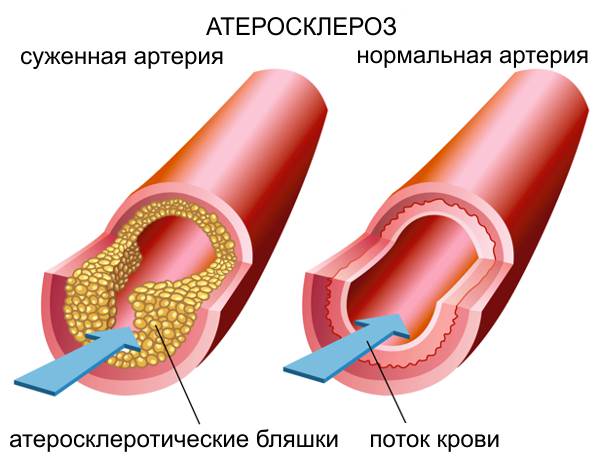
- angina pectoris in case of concomitant development of coronary atherosclerosis or subendocardial ischemia (characteristic pains in the heart);
- bacterial endocarditis in case of concomitant development of mitral insufficiency;
- development of the heart hump due to hypertrophy and dilatation of the right ventricle.
Also, there is often a high susceptibility of patients with mitral stenosis to the development of bronchitis and pneumonia.
Signs of the development of right ventricular failure
The characteristic signs of pathology include:
- heaviness in the abdomen;
- peripheral edema;
- swelling of the veins in the neck;
- ascites (accumulation of fluid in the abdominal cavity);
- hydrothorax (accumulation of fluid in the pleural region).
Causes
Mitral stenosis is also called an acquired form of heart disease.
 Causes of pathology:
- Age from 40 to 60 years (the development of the disease can begin from 20 years).
- Infectious endocarditis.
- Atherosclerosis.
- Syphilis.
- Mechanical damage to the heart.
- Severe form of calcification of the annulus and leaflets of the mitral valve.
- Myxoma of the left atrium (benign tumor).
- Congenital heart defect.
- The formation of blood clots inside the heart organ.
- Operations on the mitral valve.
- aortic insufficiency.
It has also been repeatedly observed that the disease is much more common in females.
The development of mitral stenosis can cause pulmonary hypertension in the following cases:- distribution of pressure from the left atrium to the pulmonary veins;
- increased pressure in the pulmonary veins and subsequent spasm of the pulmonary arterioles;
- swelling of the walls of the pulmonary blood vessels;
- fusion of pulmonary blood vessels.
The development of mitral valve stenosis can proceed for 30 years without showing characteristic symptoms. Only after a long period of time, the disease forms a classic clinical picture of pathology.
Diagnostics
 The main methods for diagnosing mitral valve stenosis:
- Palpation.
- Test using artificial load.
- General blood analysis.
- General urine analysis.
- Blood chemistry.
- Measurement of blood clotting.
- Phonocardiography.
- Electrocardiography (ECG).
- 24-hour Holter electrocardiogram monitoring (HMECG).
- Ultrasound examination (ultrasound).
- Echocardiography.
- Transesophageal echocardiography.
- Coronary angiography.
- Radiography.
- Probing of the cavities of the heart.
- Determination of the ejection fraction (EF).
Also, the definition of the disease is facilitated by the identification of concomitant infectious lesions of a dental, gynecological or urological nature, since the development of chronic forms of infectious diseases can significantly complicate treatment.
For an initial examination, you must contact a therapist.
Treatment
Treatment of mitral valve stenosis is divided into:
- drug therapy;
- surgical intervention
The method of treatment depends on the general condition of the patient and the stage at which the disease was detected.
So, if a pathological narrowing of the mitral valve to a size of less than 1.6 square centimeters is detected in a woman during pregnancy, in the absence of signs of valve compensation from other segments of the cardiac organ, an abortion may be prescribed due to possible complications with a high load on the heart. . However, it is worth remembering that the choice of treatment measures also remains with the woman.
Treatment with medication
 The use of medical devices helps to prevent the development of infection, heart failure and the formation of blood clots. The therapy also reduces the risk of complications.
- Antibiotics.
- Diuretics (diuretics).
- Cardiac glycosides (used to reduce the development of heart failure).
- Beta-blockers (slow heart rate, lower blood pressure).
- Angiotensin-converting enzyme inhibitors (ACE inhibitors).
- Subcutaneous administration of heparin (prevention of thrombosis).
- Angiotensin 2 receptor antagonists (ARA 2 blockers).
- Antiaggregate and anticoagulants (prevent platelets from sticking together).
The use of drugs occurs under the supervision of the attending physician. Self-prescribing drugs or changing the dosage can lead to complications and reduce the effectiveness of therapy.
Due to possible side effects of drugs, it is necessary to monitor the general condition of the patient, in case of signs of allergy or severe symptoms of malaise, consult a doctor to reduce the dosage or replace the drug with an analogue.
Surgery
Surgical intervention in violation of blood flow is carried out at stages 2, 3 and 4 of mitral valve stenosis.
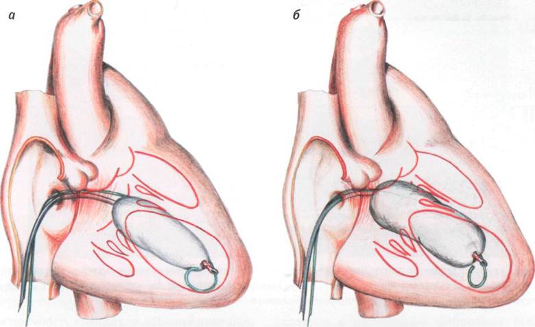
Operation types:- Balloon valvuloplasty (mechanical expansion of the heart valve).
- Open commissurotomy (separation of adhesions).
- Valve prosthetics (valve replacement with an implant of a biological or artificial nature).
 The purpose of the method of therapy depends on the form of stenosis and the presence of concomitant signs of pathology.
- narrowing of the valve ring of any scale (in the absence of calcification of the valve leaflets, the formation of blood clots inside the left atrium, with stenosis without pronounced clinical signs);
- if the pathology is accompanied by the development of atrial fibrillation;
- the absence of manifestations of mitral regurgitation after ultrasound;
- to the absence of heart disease, coronary disease (bypass surgery).
The method refers to operations with minimal pain, the duration is up to 2 hours. The operation is accompanied by the use of sedative drugs.
Features of an open commissurotomy:- the presence of signs prohibiting the use of the previous method;
- is prescribed in case of stenosis of 2,3 or 4 degrees;
- surgery is performed under general anesthesia;
- valve incision is required for the operation
- used for significant damage to the valve leaflets;
- mechanical grafts or porcine heart grafts are used during the procedure.
Medicine continues to develop not only in the direction of pharmaceuticals, but also along the path of more effective surgical intervention.
To get acquainted with the possible methods of operations of clinics, you should consult with your doctor.
Possible Complications
 If the diagnosis and subsequent treatment of the disease is delayed, there is a high probability of developing complications associated with damage to the mitral valve and other segments of the cardiac organ.
- Thromboembolism.
- Pulmonary edema.
- Bleeding in the lungs.
- Acute form of heart failure.
- cardiac asthma.
- Increased pressure in the pulmonary artery.
- Enlargement of the pulmonary artery.
Complications adversely affect further treatment.
You can also separately highlight the complications that occur after the operation.
These include:- the development of inflammation of the native or transplanted mitral valve due to infection;
- thromboembolism during the work of a mechanical graft (the exit of blood clots into the bloodstream);
- malfunction of the artificial valve.
Forecast and prevention
 Main predictions:
- In the case of the natural course of the disease, the survival of patients in the period of 5 years is 50%.
- After the operation and complex treatment with drugs, the number of surviving patients in the period of 5 years rises to 95%.
- The development of restenosis within 10 years after the operation is observed in 30% of patients.
If restenosis develops, the patient is scheduled for mitral recommissurotomy.
Measures to prevent recurrent mitral valve stenosis include measures to diagnose the work of the cardiac organ, consultation with the attending cardiologist and reduce the risk of developing streptococcal infectious diseases.
Do not forget about walking in the fresh air and airing the premises. The supply of oxygen has a good effect on the nutrition of the heart organ and its structures. However, there is a risk of contracting serious diseases that provoke a deterioration in the condition of the heart, so it is recommended to reduce contact with carriers of tonsillitis or pneumonia to a minimum.
Lifestyle in identifying mitral stenosis
After identifying violations of the mitral valve, a certain number of rules are assigned to improve the condition and reduce the likelihood of complications during the treatment period and the patient's later life.
Important factors affecting health include:- a diet that includes essential trace elements and vitamins (low levels of unhealthy foods);
- consultation with a specialist about symptoms and general condition;
- a decrease in the high level of physical activity (the child does not take part in active games);
- compliance with the regimen of taking medications;
- night sleep at least 8 hours a day, compliance with sleep and wakefulness;
- echocardioscopy at least once a year.
The basic rules for later life after surgery or when using drug therapy include measures to reduce the load on the heart.
- highly salted foods;
- marinades;
- spicy dishes;
- excess fluid;
- products that directly affect blood clotting;
- fried foods and smoked meats.
The list of prohibited and permitted products and their effect on the human body can be clarified with the attending physician during the preparation of an individual diet.
To improve overall well-being, it is necessary to adhere to the rules of a healthy lifestyle and timely undergo a diagnosis of the cardiovascular system.