Symptoms in Children of WPW or Wolff Parkinson White Syndrome
Content
ERW syndrome (Wolf, Parkinson, White) is a congenital cardiac pathology characterized by the presence of additional muscle fibers (Kent's bundle). Moving along these fibers, the impulse deviates from the correct path and prematurely causes excitation of the ventricles. The disease was described in detail in 1930. The hereditary nature of the pathology, for which the mutated gene is responsible, has been established. WPW syndrome is more often found in the male part of the population (in 70% of cases).
The mechanism of development and causes of Wolff-Parkinson-White syndrome

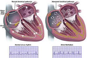
Normally, the passage of an impulse from the atria to the ventricles is carried out sequentially along a certain path.
The process looks like this:- the sinus node located in the right atrium is the starting point of the electrical impulse;
- passing through the atria, the excitation passes to the atrioventricular node (AV);
- from the AV, the impulse follows to the bundle of His, which, in turn, consists of two branches directed to both ventricles;
- and finally, along the Purkinje fibers, excitation reaches the ventricles.
Wolff-Parkinson-White syndrome is also called premature ventricular excitation due to the passage of an impulse directly from the atria to the ventricles, bypassing the atrioventricular node:Passing this path, the nerve impulse creates the conditions for an effective and synchronous contraction of the heart.
- the impulse originates, as expected, at the point of the sinus node;
- then it follows an atypical path - through the bundles of Kent or additional atrioventricular fibers - causing early excitation of the ventricles;
- a little later, the excitation of the ventricles occurs with an impulse that has passed along the correct path.
There is also the possibility of a nerve impulse returning along the same path, which causes arrhythmias.
 The detection of a dangerous pathology - Wolff-Parkinson-White syndrome - most often occurs by means of an ECG by chance. This is due to the fact that either the disease is asymptomatic, or its manifestations are similar to those of other heart diseases.
The pathology is congenital. SVC syndrome is the consequences of incomplete cardiogenesis in a child during pregnancy. During embryonic development, all humans have an additional muscular pathway from the atria to the ventricles. In the second half of prenatal development, it should completely atrophy. But due to the violation of the process, not all babies do this.
The reasons for the existence of the syndrome are:- hereditary factor. If there were cases of ERW syndrome in the family, then relatives are also likely to get sick.
- The pregnancy proceeded under the influence of negative factors. For example, the expectant mother smoked, took drugs or was addicted to alcohol. Severe stress of the expectant mother can also provoke an anomaly of the heart.
Most often, the disease is accompanied by other cardiac pathologies (in 30% of cases). Among them are heart defects, defects of the cardiac septa, connective tissue dysplasia, cardiomyopathy.
Disease classification
Depending on the external manifestations, the WPW syndrome and the WPW phenomenon are distinguished. The WPW phenomenon also shows an atypical path of the cardiac impulse, but only with the help of an ECG. There are no symptoms of this. The SVC phenomenon does not require treatment, but it is necessary to check with a doctor periodically, because the pathology can give symptoms of the syndrome. The reason for this may be increased physical activity, severe stress, alcoholism. In addition, the ERW phenomenon gives a small mortality rate (0.3%).
 Wolff-Parkinson-White syndrome can occur in several ways:
- Additional muscle atrioventricular fibers are involved in the process. They may have different directions.
- With the participation of specific muscle fibers of Kent. They are formed from tissue similar to that of the AV node. The bundles of Kent differ in their location in the heart - they can enter the right branch of the bundle of His or into the myocardium of the right ventricle.
The syndrome is also classified according to clinical manifestations.
Depending on this, the following forms of pathology are distinguished:- manifesting - this form is characterized by constant symptoms and the presence of delta waves, sinus rhythm and AV tachycardia in the ECG;
- intermittent - in this case, the signs indicated in the manifesting form appear periodically;
- hidden - the impulse travels along the additional AV connection; while the ECG will not show pathological changes; possible episodic manifestation of tachycardia.
According to the type of location of additional fibers, the disease can proceed according to one of three types. Type A involves the left-sided passage of fibers - between the left atrium and the left ventricle. Type B is characterized by right-sided passage of the impulse - between the right atrium and the right ventricle. Rarely, but it happens that one person has both types of fibers. This combination is called a mixed type.
Symptoms of ERW Syndrome
 The disease is most often diagnosed at the age of 10-20 years. The syndrome is manifested by attacks of tachycardia, and the heart rate can reach 360 beats / min. Paroxysms (attacks) begin when the impulse is first conducted through the AV node, and then in the opposite direction through the fibers of Kent or additional nodes. Because of this, the heart rate almost doubles, and blood pressure drops. The paroxysm lasts several minutes or hours. It can stop suddenly and on its own or when performing some arresting measures.
Its occurrence can be provoked by excessive physical activity, strong excitement, alcohol consumption in large quantities, pregnancy. Paroxysmal tachycardia can also occur for no reason.
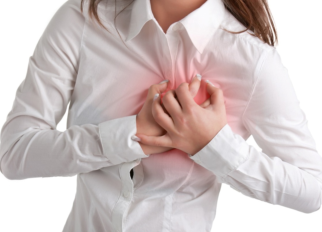
The attack is accompanied by the manifestation of other symptoms:- pulling pain or discomfort in the heart;
- dizziness;
- noise in ears;
- weakness;
- increased sweating;
- feeling of lack of air;
- blue nasolabial triangle and fingertips.
In children and adolescents, SVC syndrome manifests itself in the same way as in adults. Cases of tachycardia in a child under three years of age lead to acute heart failure.
The danger in SVC syndrome is a tendency to atrial flutter and fibrillation. This combination can lead to death.
 The disease can proceed in different ways, so there are several degrees of severity:
- With a latent course, which is observed in 40% of patients, there are no manifestations of the disease at all. It is diagnosed only by means of an ECG.
- Mild degree is characterized by rare and short bouts of palpitations.
- With moderate severity, the attacks become prolonged and can reach three hours. To eliminate them, you need to take drugs.
- In severe forms of the SVC syndrome, strong prolonged (more than 3 hours) attacks are observed, which lead to extrasystole, atrial flutter. With severe manifestations, drugs do not help. Surgery is required to prevent death. Such a manifestation of the SVC syndrome leads to disability of the second group.
If the patient is diagnosed with the SVC phenomenon, then if symptoms occur, the diagnosis of SVC syndrome is made. If up to 20 years the pathology does not manifest itself in any way, then the likelihood of symptoms of the disease is reduced to zero.
Diagnosis of ERW Syndrome
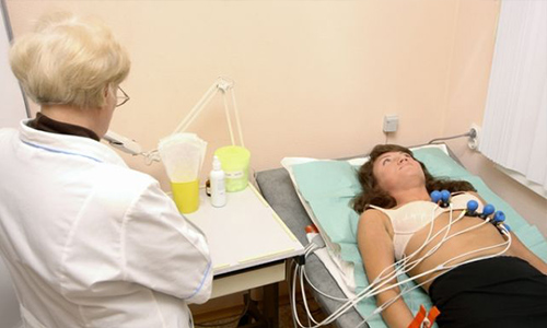
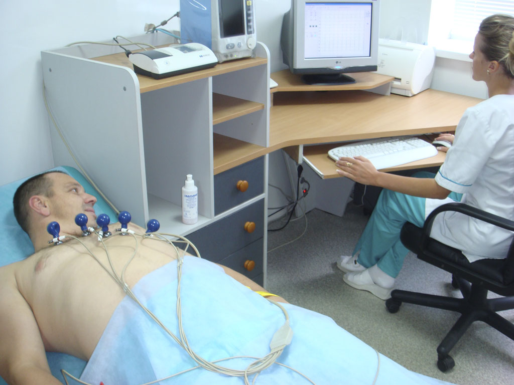
Diagnosis of the syndrome begins with an examination of the patient. The first sign confirming the disease is an irregular heartbeat when listening. Heart rate measurement completes the picture. The next step is a twelve-lead ECG.
 Examination using an electrocardiogram shows the following signs of pathology:
- a short, compared with the norm, PQ interval, which means the passage of the impulse along an atypical path;
- the presence of a delta wave, which indicates a faster, compared with the norm, excitation of the ventricles; the wavelength is directly proportional to the speed of the pulse through the additional fibers;
- a change in the QRS complex, which indicates a blockade of the His bundle of type A or type B;
- arrhythmias.
With an intermittent form of the syndrome, the patient is prescribed Holter monitoring. This is the same electrocardiography, but carried out during the day. It is also prescribed to differentiate the syndrome and the phenomenon of SVC. If during the examination signs of arrhythmia (consecutive extrasystoles) are recorded, then the patient is diagnosed with SVC syndrome, even if there are no symptomatic manifestations. If the monitoring did not show any deviations, and the patient does not feel the manifestations of the disease, then his diagnosis is the SVC phenomenon.
If the diagnosis is still not confirmed, an examination with the help of transesophageal pacing is prescribed. This method allows you to diagnose the syndrome with one hundred percent probability. Its essence is that electrodes are pulled through the esophagus to the heart and with the help of electrical impulses they make it contract in a certain rhythm. After reaching the limit of one hundred and fifty beats, the work of an additional muscle bundle stops, which indicates its presence. Also, this method is used to eliminate paroxysmal tachycardia in critical cases when medications do not help.
 The following methods are used for this:
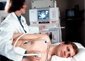
- Ultrasound is prescribed to determine the presence of concomitant cardiac disorders, which are often present in the syndrome.
- EPS (electrophysiological examination) is an invasive method that involves the introduction of catheters into the veins through the muscles to determine the location and number of additional muscle bundles. This diagnostic method is used in exceptional cases, as it has a number of side effects. Most often, EFI is indicated before surgery for the syndrome.
The presence of a diagnosis of SVC syndrome in both children and adults should warn them against excessive physical exertion (for example, big sports). Such patients should be registered with an arrhythmologist and periodically examined by him. Following the recommendations of the doctor will help to avoid complications and sudden death.
Even in the absence of manifestations of the disease, you should be attentive to your health. The syndrome is insidious in its suddenness. With a combination of favorable circumstances for its development, it can cause sudden cardiac arrest.
Diagnosis of the disease in young people of military age gives exemption from military service.
Treatment of ERW syndrome
Treatment is prescribed based on the results of the examination. If the patient has a latent form of the disease, then drugs are not required, but an annual check and prevention are necessary.
With a mild course of the syndrome, you need to know how to stop tachycardia attacks. It can be either certain physical manipulations or antiarrhythmic drugs. Techniques that allow you to cope with an attack of palpitations are called vagal. The principle of operation of these techniques is based on the activation of the vagus nerve. There are two types of nerve fibers in the body that, when attached to the heart muscle, either activate it (sympathetic fibers) or slow it down (parasympathetic fibers). The removal of an attack of tachycardia occurs through the forced inclusion in the work of the parasympathetic type of nerve fibers, or rather the vagus nerve.
 The technique for performing vagal techniques should be known to all patients with a diagnosis of SVC syndrome:
- Valsalva test. You need to take a deep breath and hold your breath in tension.
- Rinse with cold water while holding your breath.
- Mueller test. In this case, you need to try to inhale, holding your nose.
- Massage the sinus node in the neck.
- Ashner reflex. It is necessary to press on the eyeballs for 20-30 seconds.
If these manipulations did not lead to an improvement in the condition, then medication is necessary.
You can use one of the following medicines:- blockers; for example, Propranolol is a rather weak drug, it stops the manifestations of tachycardia by 50-60%, it cannot be used for low blood pressure and asthma;
- intravenous drug Procainamide - effectively relieves an attack, but is administered only in a hospital with monitoring of the patient's condition; blood pressure and heart rate are subject to control;
- Propafenone is also a very effective tachycardia drug, available in a convenient tablet form; however, it should not be used in severe cardiac lesions and is contraindicated in children and adolescents under 18 years of age.
It is also worth remembering about drugs that can not be used in the ERW syndrome. These include calcium channel blockers (Verapamil) and ATP (Adenosine). They can cause dangerous conditions of ventricular fibrillation and atrial flutter. When such processes occur, defibrillation is performed to restore a normal rhythm.
Surgical removal of pathological bundles
 The ERW syndrome forces you to take antiarrhythmic drugs constantly and for life. These drugs have many significant side effects, so more and more patients agree to surgical treatment.
- severe cases of the syndrome with prolonged and frequent attacks;
- when seizures do not respond well to therapy;
- when among the relatives of the patient there were already cases of death from the syndrome;
- when illness is an obstacle in professional activity.
The most effective method for removing an additional beam is the method of radiofrequency ablation (cauterization). Before the operation, the patient undergoes a thorough examination, and a few days before it, you need to stop taking antiarrhythmic drugs, clean the intestines a day before and do not eat. No other preparation is required.
There are also contraindications for cauterization. These include severe forms of heart failure and a recent myocardial infarction.
The course of the operation is as follows:- Local anesthesia. A catheter is brought to the removed bundle through the femoral artery (or vein), controlling the process with the help of an X-ray machine.
- This place is cauterized with an electrode using a radiofrequency pulse, and the catheter is removed.
Reviews of patients with ERW syndrome show the importance of three components for a successful cure for this disease: an experienced specialist, a comprehensive examination and an operation using radiofrequency ablation. Even with a good doctor, sometimes the diagnosis is clarified only after the operation.
Most often, the pathology is eliminated forever, but there are relapses due to incomplete removal. Complications after surgery are extremely rare (in 1% of cases). During the supply of the catheter, microtrauma of the arteries, veins and adjacent healthy parts of the heart is possible. The operation can provoke hematomas, thrombosis, spasms of the coronary vessels and atrioventricular blockade. To minimize or eliminate side effects, you should carefully choose your doctor and follow all his instructions.
The prognosis for an asymptomatic case of SVC syndrome is favorable. It is only important to see a doctor and lead a healthy lifestyle. If symptoms of tachycardia are present, then treatment or surgery is necessary. In advanced cases, the disease causes heart failure or death.