3 reasons for perfusion myocardial scintigraphy
Content
Heart disease often develops without visible symptoms. The patient learns about a severe form, a mortal threat too late, if the disease is not detected in time. Therefore, methods for diagnosing the heart and coronary arteries include informative techniques, for example, myocardial scintigraphy (SM).
The mechanism of the method and the purpose of diagnosis

Myocardial perfusion scintigraphy with stress is a diagnostic method, the purpose of which is to clarify the diagnosis. It is done after passing the ECG and ultrasound of the heart, when the diagnosis cannot be confirmed or refuted. The scan evaluates medical therapy for heart disease.
Radionuclide preparations (radioactive labels):The study is based on the principle of action of radiopharmaceuticals. Scintigraphy refers to radioisotope functional diagnostics of nuclear medicine. Radioactive isotopes are injected intravenously into the human body, participate in the bloodstream, reach the place of study - the heart.
- Tc-99m-MIBI;
- thallium-201;
- therofosmin labeled with technentium-99.
Perfusion is the infusion of solutions and substances into the vascular system of the body. The same under this term understand the natural blood supply to tissues. The SM method is non-invasive, meaning it does not require surgical incisions.
During the test, the person is exposed to stress. This is done in order to cause the activity of the heart, to create conditions for angina pectoris. Supernatural efforts are not required from a person, all exercises correspond to physical capabilities. Voltage conditions are as close as possible to the natural conditions of human life. SM with a load becomes an independent study, or is part of a comprehensive study.
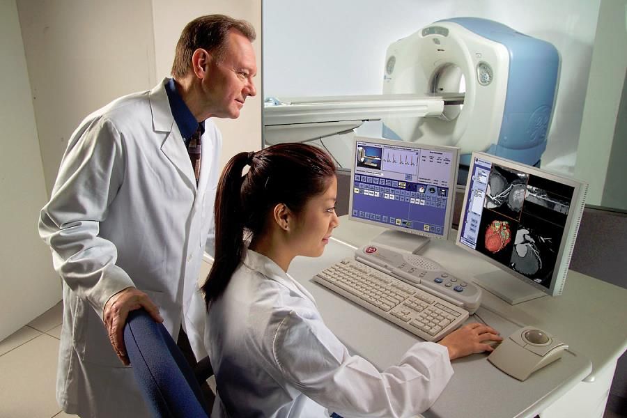
 The result of scintigraphy is a picture of the heart in different projection axes with highlighted areas. With the help of a radiometric apparatus, you can see the gamma radiation of radioisotopes on the screen, analyze the state of blood circulation in all parts of the heart muscle.
- stenosis of the arteries;
- ischemia;
- consequences of a heart attack (tissue necrosis);
- pathological changes associated with defects.
The main purpose of scintigraphy is to identify deviations from the normal movement of blood, to detect obstacles that have arisen as a result of damage to the myocardium and coronary arteries.
Research using radioactive substances is harmless to health. Produced in special radiological research centers. Radiation exposure is too low because radiopharmaceuticals quickly and completely decay.
Basis for diagnosis
Myocardial scanning is performed if the patient:
- complains of angina attacks, and she takes on a permanent character;
- suffered a heart attack;
- undergoing rehabilitation therapy after a heart attack or heart surgery.
Perfusion scanning determines the likelihood of complications after a heart attack, the localization and size of the area of affected tissues. Necrotic areas are not supplied with blood, so the radiotracer cannot get into the affected area. A specialist in dim gamma radiation in the picture finds and fixes areas with necrosis.
 By the same principle, stenotic lumens of the coronary arteries are found, due to which IHD (ischemic heart disease) occurs. Through the narrowing, blood passes poorly, oxygen and blood starvation of the organ occurs. Therefore, patients at risk for coronary artery disease are recommended to undergo an examination using radionuclides.
- diabetes, hypertension;
- smoking experience more than 20 years;
- cases of heart disease among relatives (heredity);
- age group from 45;
- high cholesterol.
For example, angina is caused by atherosclerotic processes, heart attack and diseases of the gastrointestinal tract. The true cause of chest pain is established during the study.
Scintigraphy data are the basis for revascularization when they confirm:- ineffectiveness of medical treatment of angina pectoris, heart attack;
- the presence of fragments of the myocardium, where it is possible to restore blood supply.
Diagnosis establishes the feasibility of surgical intervention, and after the operation helps to track the dynamics.
 Contraindications to the study:
- pregnancy, breastfeeding;
- allergy to radiopharmaceuticals;
- acute heart attack;
- unstable angina attacks;
- valvular defects;
- critical forms of arrhythmia (fibrillation, hypotension, hypertensive crisis).
- patients weighing more than 140 kilograms;
- with endocarditis, myocarditis, pericarditis;
- in the acute period of stroke, thromboembolism.
With these contraindications, the study is not carried out due to the general deterioration in the patient's health. The weight and volume of the body will interfere with the examination in the tomograph if they exceed the allowable values (the person may not fit in it). Large breasts are also the cause of errors.
Getting results and decryption
The technique of scintigraphy includes two stages:
- Study at rest.
- Examination after exercise or administration of a drug that activates the work of the heart.
In time, both stages take about four hours.
 At rest, the gamma camera records the parameters of the circulation of the radioactive drug with the blood. A series of shots gives an image of the heart in different planes as the camera moves around the patient. It remains motionless during the scan.
After the decay and removal of the substance from the body (the next day), the patient undergoes the second stage of the study. The purpose of the stage is to monitor myocardial blood supply with an increase in heart rate.
Ways to increase heart rate:- load in the form of physical exercises;
- the introduction of adenosine (intravenously administered, simulates the load).
Exercises are performed on a treadmill (treadmill), bicycle (veloergometry). However, if increased physical activity is contraindicated, medical toning of the heart muscle is used.
In both cases, the activity of the heart is monitored using an ECG. The results are taken when the heart rate has reached a certain level (an acceptable indicator for the patient) or the symptoms of the pathology have appeared, that is, angina pectoris with coronary artery disease.
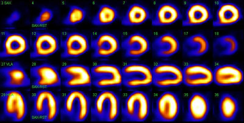
The resulting images are compared after 4 hours. Spots of cold and warm shades indicate the amount of accumulated radiopharmaceutical. The indicator of the norm is the uniform distribution of the substance over the myocardium.
- Fully reversible (the color of the area is completely restored).
- Partially reversible (color is partially restored).
- Persistent (does not recover, angina pectoris does not go away).
 Technetium accumulates in the affected tissues. Pictures of a healthy person before and after exercise will not differ from each other. The substance will not appear on the screen as a red spot. The entire surface to be examined will be cold in color. A persistent accumulation defect indicates a heart attack, cardiosclerosis, and indicates the presence of post-infarction scar tissue.
Thalium accumulates in healthy tissues. To decipher the defect, cold-colored spots are analyzed.
The accumulation defect indicates a violation of perfusion. The myocardium is divided into segments into which blood enters along with a radioactive label.
The degree of accumulation is measured as a percentage:
- Normal perfusion is 75% or more of the maximum blood supply.
- Moderate - 51-74%.
- Reduced perfusion - 30-50%.
- Decreased perfusion is expressed - 30%.
The percentage is converted into points. A four-point system is used, where normal perfusion is estimated at 0 points, and a pronounced decrease in blood circulation is estimated at 3 points. The higher the percentage of perfusion or the lower the score, the more viable the myocardial site. Doctors see an opportunity to restore blood circulation in him.
When interpreting, the location of the segment of the walls of the left ventricle with impaired perfusion is indicated: lower, lateral, anterior walls, interventricular septum.
Preparation of the subject
 For a successful and accurate diagnosis, the patient must:
- Get directions.
- Choose a clinic.
- Stop taking heart medication.
- Refrain from eating and drinking on the day of the test, and exclude foods high in caffeine (drinks, chocolate, etc.) from the diet a few days in advance.
- Women get an ultrasound to check for pregnancy.
- Provide an insurance policy, a medical card with notes on the surgeries.
In the direction, the attending physician must indicate the diagnosis and symptoms. The results of other examinations (stress electrocardiogram), prescriptions, medicines attached to the referral will help clarify the diagnosis.
The action of drugs leads to errors:
- Curantyl;
- beta-blockers of adrenaline and calcium channels;
- nitrates;
- Methylxatin;
- Pentoxifylline.
Refusal of medicines occurs with the permission of the doctor on average two days before the test.
The clinic can provide everything you need, but for personal comfort it is better to have with you:
- a set of change of clothes and shoes;
- towel;
- foods high in calories (for about two meals).
Nursing mothers are contraindicated in the SM procedure. In case of an urgent need for examination, experts recommend expressing milk on the day of the test, within a day after it.
Possible complications and side effects
 The examination is safe for humans. Slight discomfort occurs when drugs are administered intravenously, and the subject is required to lie still in the gamma chamber for a long time.
The examination uses radiopharmaceuticals. They are distinguished by low radioactive radiation, they are excreted from the body within two days. Cause allergies less frequently than X-ray contrast agents. But the dose of radionuclides is enough to harm the embryo during pregnancy.
Individual intolerance can manifest itself in the form of:- manifestations of skin allergic reactions (rash, redness);
- edema;
- failures of arterial pressure;
- nausea;
- frequent urge to urinate.
After the examination, drinking plenty of water accelerates the excretion of isotopes from the body, relieves the manifestation of the listed symptoms, and reduces the radioactive load.
During exercise, deterioration, dizziness, weakness may occur. Rapid fatigue is accompanied by shortness of breath and chest pain, so it is necessary to interrupt the study. A similar drug load will cause similar sensations and hot flashes.
Cost of diagnostics
SM has become widespread in Europe and America.
In the CIS countries, anyone who wants to be examined is prevented by:- expensive and rare equipment;
- lack of qualified specialists;
- service cost.
 Perfusion scanning requires a gamma camera, specialists in this type of diagnostics, and experience in organizing and conducting scans.
- RNIMU them. N. I. Pirogova (Moscow).
- FSBI "Clinical Hospital" of the Administration of the President of the Russian Federation (Moscow).
- Russian Scientific Center for Radiology and Surgical Technologies (St. Petersburg).
- Alexander and Elizavetinskaya hospitals (St. Petersburg).
- FSBI "NMITs them. V. A. Almazov» of the Ministry of Health of Russia (St. Petersburg).
In small towns, the service is often not available in state institutions, so patients turn to private medical centers.
The price of perfusion scanning at rest varies from 4,000 to 7,000 rubles. Carrying out the SM complex at rest and with a load will be 13,000-15,000 rubles.
In the price list you can find the following name: "Myocardial scintigraphy and SPECT with Tc-pyrophosphate". SPECT (or SPECT) is single photon emission computed tomography. This is one of the methods of scintigraphy. The patient should not be afraid of the wording of another scanning option - PET (positron emission tomography). The listed methods are used separately and combined at different diagnostic sites in order to increase the information content of the scinogram.
The risk group for coronary heart disease becomes younger over the years. Cases of heart attack and stroke are not always associated with old age. Accurate diagnosis is the first path to timely treatment.