Aortic aneurysm of the heart
Content
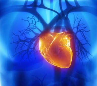
Aortic aneurysm of the heart, what is it and how to treat it is a common question after diagnosis. Timely access to a doctor allows you to avoid dangerous consequences.
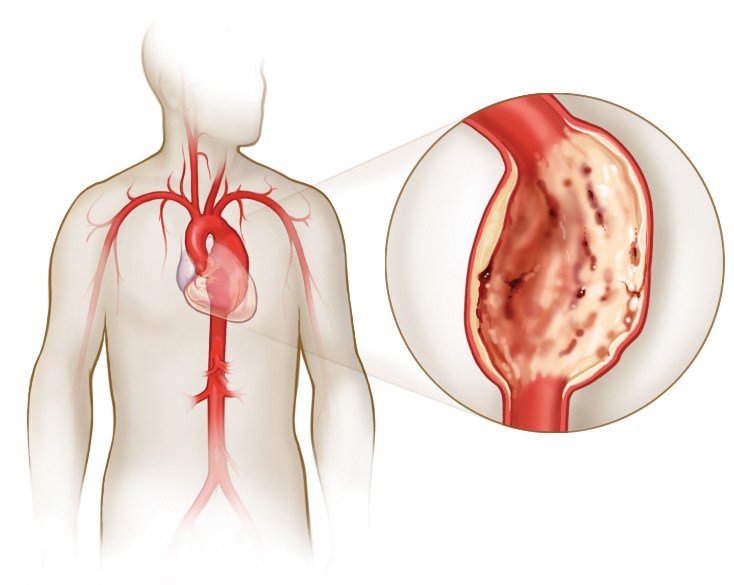
Aortic aneurysm of the heart is a pathology characterized by a local increase in the volume of the aortic vessel. It can form in any part of it, has persistent clinical manifestations. With a long course of the disease or in case of injury, a complication in the form of a dissecting aortic aneurysm is possible. Cardiologists classify the disease depending on the clinical, morphological and etiopathogenetic signs. But the fundamental criterion in the treatment is the location of the pathological expansion.
Causes of the disease

The area of unnatural expansion of the walls of the aorta, as a rule, does not exceed a size of 5 centimeters. An aneurysm is a dangerous pathology, since in the process of increasing the local volume of the artery, smaller vessels can be squeezed, which play a significant role in the "nutrition" of the heart muscles.
Aneurysm is a pathological expansion that has an irreversible process.
It develops due to dystrophic changes in the vessel.
Provoke such damage can:
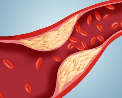
- Atherosclerosis. Most often observed in old age.
- Injury to the chest, if at the same time there was an infringement of the heart muscle.
- Inflammation of muscle fibers, when the process is chronic.
- Fibrous dysplasia of the chest.
- Heart attack. Partial death of heart muscle cells leads to the formation of a post-infarction scar, which is formed from the connective tissue.
- Arterial hypertension and other cardiovascular diseases.
- Marfan's disease with loss of elasticity of the connective tissue. It is genetic in nature and can lead to a dissecting aortic aneurysm.
Often an aneurysm is the result of insufficiency of the middle layer of the artery wall. Such a defect is often congenital. In this case, with an increase in pressure on the site, a local violation of the integrity of the wall of the aortic vessel is observed.
 In addition, the development of the pathology of the aortic vessel can provoke non-specific degenerative processes (medion necrosis) occurring in the middle layer of the arterial wall. Most often, such changes are noted in individuals with a generalized pathology of connective materials.
The acquired form of the disease is mainly a consequence of an infectious or immune disease of the aorta, which was accompanied by inflammation of its walls.
The main difference between an aortic aneurysm and similar pathologies of other arteries is that the expansion has only a fibrous composition, there are no signs of laminar blood flow at all.
Types of pathology
Aortic aneurysm is classified depending on clinical, morphological, etiopathogenetic signs and the place of its manifestation.
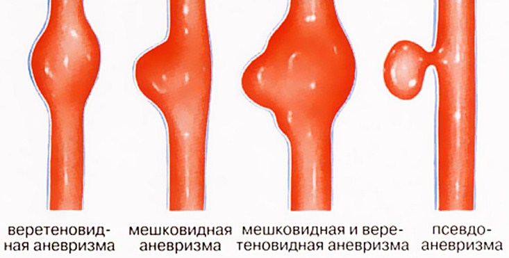
In appearance, it can be divided into the following types:
- Flat. It is on the same level with the heart, protruding into the organ area.
- Mushroom. In this case, the shape of the aneurysmal formation resembles a mushroom.
- Sacciform. On the one hand, the aneurysm is more convex than on the other.
- diffuse. In this case, the formation changes its parameters depending on the change in blood pressure.
Therefore, doctors divide this pathology into three types, which allow a more accurate diagnosis:
- So, with a true aneurysm, the manifestations completely coincide with the studies.
- The functional form of the pathology is characterized by small necrotic processes in the walls of the artery, due to which its functionality is partially or completely lost.
- With a false aneurysm, MRI readings differ from the clinical picture. In this case, adhesions or tumors are found that have nothing to do with pathology.
With regards to leakage, there are three forms of the disease:
- Acute. The most dangerous. It develops suddenly and quickly, is the result of a heart attack or inflammation. If you do not take immediate operational measures, a fatal outcome is possible.
- Subacute. Occurs after any heart disease or surgery. Has no pronounced symptoms.
- Chronic. It is distinguished by an even flow, without severe pain manifestations. With this form, thinning of the walls of the artery occurs.
Symptoms of the disease
The longer the disease proceeds, the more the bag increases in size. Therefore, diagnosis at an early stage is very important, since, otherwise, there is a very high risk of aneurysm rupture into the nearby cavity, heart sac, pulmonary trunk.
Characteristic signs of the disease:
- Aneurysm of the aortic sinuses. This pathology is accompanied by valvular insufficiency or a decrease in the lumen of the blood channels. With a large increase in such an aneurysmal sac, compression of the pulmonary trunk, right-sided ventricle and atrium occurs. This condition provokes the development of heart failure. As a result, an increase in the liver and vessels of the cervical region, the appearance of edema are noted. The rapid progression of an aneurysm of the aortic sinuses can lead to a tragic outcome.
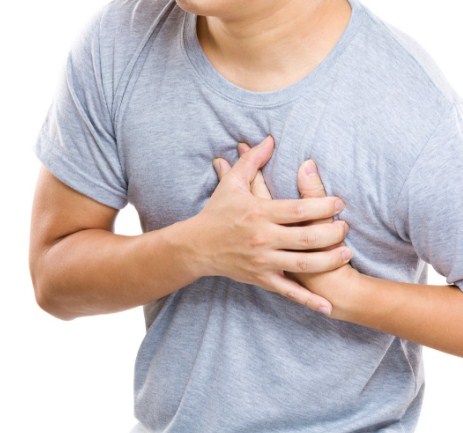
- Aneurysm of the ascending aorta. With this disease, the patient feels a dull pain in the chest, sometimes accompanied by bouts of shortness of breath. In the case of a significant increase in the aneurysmal sac, atrophy of adjacent tissues is possible. The patient feels a pulsation between the second and third ribs. If compression of the superior vena cava occurs or the formation breaks into its cavity, swelling of the neck, upper limbs, and face occurs.
- Aneurysm of the aortic arch. With this type of pathology, compression of the trachea and bronchi occurs. The patient has shortness of breath, it is difficult for him to take a breath. In case of compression of the main bronchus, lung atelectasis is possible. With an aneurysm in the aortic arch, hemoptysis is possible. Very often it precedes the rupture of the aneurysmal sac. If compression of the left lower laryngeal nerve has occurred, the patient often develops a dry cough, the timbre of the voice changes, and asthma attacks are not uncommon.
- Aneurysm of the descending aorta. An increase in the aneurysmal sac in this case leads to compression of the nerve roots. This is manifested by severe pain that cannot be eliminated with medications. Also, lower paraplegia (with pressure on the vertebrae), lung atelectasis (in case of compression of the left lung), difficulty in moving food, bleeding (with pressure on the esophagus or rupture into its cavity) may also appear.
With untimely diagnosis, the formation of a dissecting aortic aneurysm is possible. This form of pathology is manifested by a strong pain sensation in the chest, which cannot be stopped by medications and collapse. Quite often, the manifestations of this form of pathology can be confused with the symptoms of an acute heart attack.
Diagnosis and treatment
Most often, patients turn to the therapist with complaints of chest pain. The doctor collects an anamnesis, analyzes the symptoms and refers to a cardiologist.
To confirm the diagnosis, the doctor may prescribe the following measures:
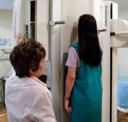
- X-ray. It is carried out in three projections. Expansion of the shadow of the vascular bundle, bulging of the aneurysm of the descending artery into the left lung, calcification of the formation is revealed.
- Ultrasonography. Determines the parameters of the aneurysmal formation, the condition of the vessels branching off from the aorta, valve defect.
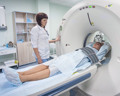
- CT scan. When the lumen of the aortic vessel is more than 4 centimeters, it is considered as an aneurysmal. In this case, the condition of large blood channels and the presence of dissection in case of exfoliating pathology are ascertained.
- Angiographic study. It is carried out before the operation to determine the nature and extent of the intervention.
Not always the diagnosis requires immediate surgical intervention. But there are some indicators that are an indisputable argument for the operation.
These criteria include:
- expansion of more than 5 centimeters;
- aneurysmal formation, which may lose integrity;
- risk of complications of thromboembolic type;
- rapid growth in education.
The technique of surgical intervention is directly dependent on the location of the pathology:
- In the case of an aneurysm of the ascending segment, the operation is performed through a median sternotomy. First, the artery is exposed and switched off from the bloodstream. For this, clamps are applied at a distance of about two centimeters from the formation. Removal of the saccular formation occurs completely. But the distance of the vascular wall of one centimeter from the clamp remains unchanged. After removal of the aneurysm, in case of a small defect, the surface is sutured; in case of a significant wound, synthetic material is sutured.
- If the aneurysm of the ascending aorta is fusiform, a technical device for cardiopulmonary bypass is used during the operation. After exposure of the vessel, a transverse clamp is applied slightly above the branch of the brachycephalic trunk. Elimination of aneurysmal formation is carried out in conjunction with the introduction of cannulas into the coronary vessels. This improves coronary perfusion. Since the fusiform pathology is of considerable size, after its removal, tissue replacement with an allograft is required.
- Elimination of an aneurysm of the aortic arch is carried out only with the use of a heart-lung machine. This area, together with the outgoing vessels, is excluded from the circulation with the help of special clamps. After removal, the site is replaced with a graft.
- Elimination of an aneurysm located in the descending aorta requires only partial use of a heart-lung machine. Vessels that “nourish” the upper body are not excluded from the circulation. A left-sided thoracotomy is performed with further opening of the pericardium. Clamps are placed across the artery. Resection of the site and suturing of the graft is carried out to the remaining part of the artery wall.
 Very often, significant aneurysm sizes lead to hemodynamic disturbances. In this case, medical correction is required. If the patient has contraindications to surgical intervention (old age, concomitant diseases in the stage of decompensation), drug treatment is attributed. In this case, antihypertensive drugs of an etiopathogenetic orientation are prescribed. In addition, it is recommended to radically change your lifestyle.
If left untreated, this disease has a very poor prognosis.
Due to rupture of the formation, heavy bleeding, hemorrhagic shock, a fatal outcome is possible.
After the operation, the patient completely changes his lifestyle. To maintain a normal life, he needs to avoid stress, great emotional and physical stress, adhere to proper nutrition, a strict daily routine, and strictly follow the doctor's instructions.