Symptoms and treatment of heart disease pentade of Fallot
Content
- 1. Place of Fallot's tetrad in the UPU system
- 2. Mechanism of hemodynamics in tetralogy of Fallot
- 3. Causes of CHD
- 4. Types and forms of Fallot's tetrad
- 5. Basis for diagnosis
- 6. Methods for diagnosing tetralogy of Fallot
- 7. Drug treatment of Fallot's tetrad
- 8. Surgical method
- 9. Living With Fallot's Thetad
The fight against congenital heart defects begins long before the baby is born. A council of doctors considers all possible risks together with the expectant mother. Often a defect makes it possible to give birth to a child, to get stronger, to wait for the moment when the strengthened organism can endure a serious operation. Such operable defects include Fallot's tetrad, which in the conditions of modern medicine has long ceased to be a death sentence.
Place of Fallot's tetrad in the UPU system

Congenital heart disease (CHD) is a pathology of development in the embryonic and postnatal period. This may be a delay in the formation of an organ, an abnormal structure, disorders of cardiac activity (circulation). Half of children with CHD die before the age of one year, in the first week of life - 20%. If it was possible to overcome the most dangerous first year of life, then without an operable solution to the defect, the child will not live older than 12 years.
According to the Taussig classification, two groups of CHD are distinguished:Tetralogy of Fallot ranks third among the most common heart defects - up to 13% of all CHD, according to various sources. The most common are atrial septal defects (ASD) and ventricular septal defects (VSD).
- white (arterial and venous blood does not mix);
- blue (arterial and venous blood is mixed).
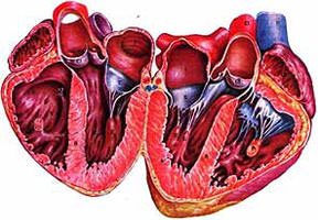
CHD tetralogy of Fallot - a "blue" defect with abnormal vascular discharge, normal position of the ventricles.
It is a complex of four (therefore "tetrad") pathologies:- VSD;
- stenosis of the lumen of the pulmonary trunk;
- right ventricular hypertrophy;
- dextroposition (displacement) of the aorta.
The defect occupies a place between the triad (without displacement of the aorta) and Fallot's pentad (all signs of a tetrad and ASD).
The vice system is named after the French pathologist ELA Fallot (1850-1911). At the turn of the nineteenth and twentieth centuries, pathology was considered incurable, discovered only after an autopsy.
Life expectancy with a diagnosis of Fallot's tetrad directly depends on the degree of heart failure. The operation in the first year of life gives a chance to save the child.
The mechanism of hemodynamics in tetralogy of Fallot
 Cardiac activity is the basis of blood circulation, which, in turn, provides metabolic processes and oxygen access to cells. Blood during normal heart function should freely enter the aorta after being enriched with oxygen in the lungs.
- VSD is that the septum between the ventricles is either absent altogether or seriously deformed;
- stenosis means narrowing of the pulmonary valve.
As a result, arterial blood mixes with venous blood at the point where the septum should separate the two ventricles. A narrow passage in the pulmonary artery does not allow the heart to throw blood into the lungs. There is stagnation of venous blood.
Blood with a sufficient level of oxygen does not enter the organs and tissues. Hence the name of the classification of such defects - "blue". As a result of hypoxia (oxygen starvation), a blue tint of the limbs appears. This blue discoloration of the skin and mucous membranes is called cyanosis.
The degree of hemodynamic disturbances is determined by the severity of the defect, that is, by how much the septum is deformed and how narrow the passage from the right ventricle is:- a significant narrowing of the lumen of the pulmonary artery, the absence of a lumen, the occurrence of an obstacle (most of the mixed blood goes into the aorta);
- moderate stenosis allows blood to pass, which turns the "blue" malformation of Fallot into "white" (no cyanosis);
- cyanotic type of defect, in which the blue of the limbs occurs due to narrowing.
In the first case, the blood, in order to enter the pulmonary circulation, enters the lateral branches of the blood flow (collaterals), bypassing the main blood trunk.
A heart affected by a defect cannot fully perform its functions. All body systems suffer from this, so a surgical solution is needed to restore oxygen supply.
Causes of CHD
 In the first trimester of pregnancy, the main organs of the fetus appear. From the second to the eighth week, the heart is formed.
Abnormal development of the heart begins with the fact that the aorta is in the wrong position. Further, the pathology works in a chain: the position of the pulmonary trunk shifts, it stretches and becomes narrower, the septum of the arterial cone does not connect to the septum of the ventricles, an incomplete septum (defect) appears, the right ventricle expands.
Tetralogy of Fallot occurs when a pregnant woman:- uses drugs and alcohol;
- abuses smoking;
- works in dangerous and harmful working conditions;
- at the household level, it comes into contact with toxic substances, paints, chemicals;
- in the first trimester she had rubella, influenza, scarlet fever, suffered measles;
- has relatives with operated CHD, operated on herself;
- took sedatives, sleeping pills and other medicines;
- underwent a course of hormonal therapy.
In the case of rubella, the pregnant woman receives a recommendation from doctors to terminate the pregnancy at an early stage.
The mother herself can start the process of improper development of the main organ of the unborn child if she does not find out about her pregnancy in time.
Types and forms of Fallot's tetrad
 The classification of Fallot's tetrad is based on the degree of changes in the structure of the heart.
- Embryological.
- Hypertrophied.
- tubular.
- Multicomponent.
The embryological type means that the septum is displaced forward, to the left and down, the narrowing corresponds to the muscular ring of the pulmonary valve, the valve itself is not changed, or moderate hypoplasia (underdevelopment) is observed.
Each subsequent type of defect is complicated by pathology. For example, the hypertrophied type of Fallot's tetrad indicates that hypertrophy of the septum is added to the characteristics of the embryological type. This means that it is displaced and increased in volume and mass.
The multicomponent type is the presence of all pathologies and the elongation of the septum.
There is also a classification that is based on the characteristics of blood circulation.
There are forms of Fallot's tetrad:- With fusion (atresia) of the entrance of the pulmonary artery.
- Cyanotic ("blue").
- Acynotic ("white").
With the growth of deviations in the development of the heart, the patient's condition worsens. The transition from the acinotic form to severe cyanosis indicates a progressive defect.
Basis for diagnosis
 The manifestation of the main symptom - cyanosis - occurs in five forms:
- Acinotic (no cyanosis).
- Late cyanotic (cyanosis appears at 6-10 years).
- Severe cyanotic (cyanosis is manifested by attacks accompanied by shortness of breath).
- Classical cyanotic (cyanosis appears at 2-3 years).
- Early cyanotic (cyanosis manifests itself in the first year of a child's life, especially from the second to fourth months).
In four out of five forms, cyanosis becomes a visible symptom of Fallot's tetralogy. "White" form means the normal color of the skin. In other cases, the nasolabial triangle, hands, feet, and upper body turn blue. The blue tone of the skin intensifies when the baby cries, tenses up, and may appear during feeding.
The remaining symptoms of the defect are detected both during physical activity and at rest:- shortness of breath when feeding, walking, running;
- severe weakness, increased weakness;
- physical and motor underdevelopment;
- thickening of the fingers (reminiscent of "drumsticks").
Seizures are usually caused by acute hypoxia and maximum ejection of blood through the deformed wall. The frequency of acute manifestations of symptoms depends on how narrowed the pulmonary artery is: the more pronounced the stenosis, the more often the child suffers from seizures.
 The defect interferes with the normal development of the child, therefore, over time, they say about its presence:
- late growth spurts (with a delay, the child learns to crawl, sit, walk);
- weight loss;
- "heart hump" or flat chest;
- rachiocampsis;
- an increase in the distance between the teeth (grow incorrectly);
- a tendency to be flat-footed.
Particularly dangerous attacks of shortness of breath and blue in the face. They last half a minute, sometimes five minutes, but always entail danger, as they can end in a stroke or death.
To help the child survive the attack and stop it in time will help:- the position "squatting" and "knee-elbow" (this way you can reduce the load on the heart);
- oxygen mask;
- administration of medications that reduce tachycardia;
- morphine (reduces breathing rate, anesthetizes).
An attack is a reason to call for specialized help. The half-hour duration of the attack indicates the need for an urgent operation.
Methods for diagnosing tetralogy of Fallot
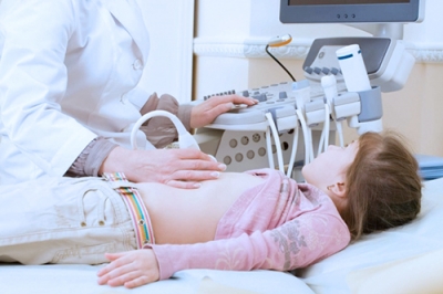
Signs of Fallot's tetrad are most clearly seen on an ultrasound of a pregnant woman during the third trimester, when the organs are all formed and their correct or incorrect activity is visible. Before this period, a high-quality ultrasound diagnostic device will detect deviations for up to 22 weeks.
 A woman will find out the predisposition in percentage terms at the first screening as part of a genetic study. Geneticists will conduct a detailed analysis for chromosomal diseases.
- Weakening of the II tone, pronounced noise during systole according to the results of phonocardiography.
- "Slipper" or "Dutch boot" on ultrasound of the chest.
- Deviation of the electrical axis of the heart to the right, myocardial hypertrophy, incomplete blockade of the His bundle on the ECG.
- Anatomical defects of the defect on ultrasound of the heart.
- Violations of blood flow in the heart muscle according to dopplerometry.
- Exceeding the norm of erythrocytes twice as a result of a general blood test.
- Increased pressure in the right ventricle as a result of sounding of the cavities.
To exclude a diagnostic error, differential studies are carried out.
It is necessary to distinguish Fallot's tetrad with diseases such as:- Eisenmenger's tetrad (instead of stenosis - expansion, which is determined on x-rays);
- displacement of the main vessels, aorta and pulmonary artery (with it, the heart greatly increases in size with age);
- atresia of the tricuspid valve (occurs in the left ventricle, and not in the right, as in Fallot's tetrad);
- stenosis of the pulmonary artery (without the other components of the tetrad and the "boot" in the picture);
- univentricular heart.
The doctor can also analyze the manifestation of external symptoms, palpate the contours of the heart.
Drug treatment of Fallot's tetrad
 The only way to defeat the defect and reduce the likelihood of death from heart failure in childhood is surgery.
- stops an attack;
- prepares the body for surgery;
- supports the work of body systems when it is necessary to wait for a good time for the operation.
Newborns with a detected pathology are placed in special boxes to reduce oxygen starvation of tissues, inhalations are shown to older children. Parents at home should ensure sufficient humidity.
As part of medical care, they use:- Morphine to reduce pain;
- Phenylephrine or Ketamine for high blood pressure;
- oxygen therapy prevents oxygen deficiency (ineffective oxygen therapy);
- Prostaglandin to maintain patency of the arterial duct;
- Reopoliglyukin for active blood circulation and prevention of thrombosis;
- sodium bicarbonate restores water-electrolyte and oxygen-alkaline balance;
- glucose for myocardial nutrition;
- Eufillin to improve blood flow.
The use of drugs is an auxiliary measure. Drug therapy is carried out by qualified cardiology specialists and does not replace the only effective solution for the tetralogy of Fallot - surgery.
Surgical method
 A confirmed diagnosis of a defect means a mandatory operation. A surgical decision in the first year of life is favorable in many respects, including helping to reduce the risk of complications.
- Palliative (bypass).
- Radical.
The palliative method is often used in emergency cases, when the attack is not stopped on its own and with the help of drugs, the physical lag in development is strongly pronounced, and the manifestations of cyanosis have intensified. It consists in carrying out an intrapericardial anastomosis (connection) of the aorta and the pulmonary artery. Restoration of the circulation circle is possible through prosthetic connection, implantation or connection of the subclavian artery into one node with the pulmonary artery.
The problem of narrowing of the pulmonary valve is solved by introducing a special expansion balloon into the arterial cavity. This technique is called endovascular balloon valvuloplasty for valvular stenosis. It is done under X-ray.
Palliative operations also include plastic surgery of the right arterial cone (infundibuloplasty).
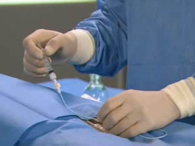
- Under general anesthesia, the chest is dissected, since the operation is performed on an open heart with the heart-lung machine turned on.
- An incision is made in the area of the right ventricle.
- Valve plasty (dissection) is performed.
- A septal defect is eliminated by applying a fragment of artificial origin or from biological material.
There is a danger of damaging the coronary arteries, hurting the pacemakers, so the operation is performed only by cardiac surgeons with high and narrow qualifications.
 The decision to operate is made by the child's mother. Patients who successfully survived such procedures share their fears of a serious step. Their revelations help to understand the safety of the method and the importance of the operation for later life:
“The diagnosis was made at 31 weeks pregnant. I was immediately sent to the perinatal department at the Bakulev Center. The doctors said that the main thing is not to add prematurity to the heart disease, so you need to “reach” a difficult pregnancy. For the operation, they chose between Bakulevka, the Filatov hospital and a variant in Germany. We chose Filatovskaya. Followed by a cardiologist for up to 6 months. The state of blood supply worsened gradually, but without visible signs. The child developed normally. Then the operation, five hours of anesthesia, resuscitation, two days later transfer to the ward, thirty days postoperative period "
Serezha's mother, Moscow
“My daughter was born with a VSD. Until the age of 19, she lived a normal life until she was diagnosed with Fallot's tetrad. Doctors predicted death in five years. And she wanted to live, family, children ... The chances were negligible, but her daughter decided on an operation. I could no longer influence the decision of an adult child. For the first time, the doctors touched something, stagnation began in the lungs. Three weeks later, a pacemaker was put in, and she got better. She received a third disability group, but this did not stop her from meeting her love, getting married and getting pregnant. At a period of 39 weeks, a caesarean section was performed, the baby was born with a healthy heart. And her daughter will have a second operation, but now it is necessary for the happy life of her family. Hope only for the best!”
Svetlana, Chelyabinsk
Women who have encountered Fallot's tetrad during pregnancy call the most important weapon against vice - awareness. It is important to know everything about the surgical decision of the CHD, about possible risks and complications, to work together with doctors and cardiologists in order to give a happy and healthy life to the child.
Living with Fallot's tetas
 Without surgery, children with Fallot's tetrad live no more than 12 years. Any infectious disease can pass with complications and lead to a sharp deterioration in the work of the heart.
If a heart with a defect allows a person to live to the age of forty, then as a high degree invalid who cannot exist without outside help.
Every patient with a defect has a chance for a physically active life. The earlier the operation is done, the faster the child recovers and comes to normal development. He successfully realizes himself in a full-fledged society. If surgical correction is carried out in an adult and fully formed person, then positive dynamics appear later.
- damage to pacemakers;
- trauma of the coronary vessels;
- inefficiency of plasty of stenosis and septum.
The prognosis of life among patients in 95% is favorable, but there are also statistics according to which people die after a radical correction. Damage to the Hiss bundle, for example, threatens with subsequent heart rhythm disturbance. From different types of arrhythmia, the patient can come to flutter and atrial fibrillation. This is due to chronic overload of the right ventricle.
 There are no visible signs of such changes, death occurs suddenly. And in the latter case, in the absence of improvements after plastic surgery, a second correction will be required.
These examples once again emphasize the importance of a competent approach to choosing a cardiology clinic.
During the period of waiting for the operation and in the postoperative period, the patient is observed by a cardiologist and a cardiac surgeon. There is also a degree of disability.
Disability is awarded in accordance with the law:- after the operation, the patient is observed and treated for another 4 months;
- the medical organization that carried out the treatment sends for a medical and social examination;
- examination reveals the presence or absence of signs of disability, analyzes the results and effectiveness of treatment;
- the degree of circulatory insufficiency and other factors are established, in accordance with which a disability group is awarded.
After two years, a recertification is required.
Thus, if the narrowing of the pulmonary artery persists in the postoperative period, blood circulation is disturbed, there are complications, then the person has the right to apply for a disability group.
The main recommendation of cardiac surgeons after surgical correction of Fallot's tetrad is observation. A child who suffered it in early childhood may not notice any consequences and develop fully. However, parents should always be vigilant about the condition of the baby, visit a cardiologist, undergo appropriate diagnostics in order to detect abnormalities in the work of the heart in time.
Tetralogy of Fallot is a congenital heart defect that has a favorable surgical solution. Gone are the days when a pregnant woman was persuaded to abandon her child or terminate her pregnancy. Life with such a diagnosis is possible if all four components of the defect pathologies are corrected in childhood. After that, you need to know everything about the disease and adhere to the rules of a healthy lifestyle in order to take care of the most important organ of the human body.