What is echocardiography of the heart - decoding
Content
Echocardiography of the heart is one of the modern diagnostic methods that allows you to accurately determine the condition of the heart and evaluate its contractile activity.
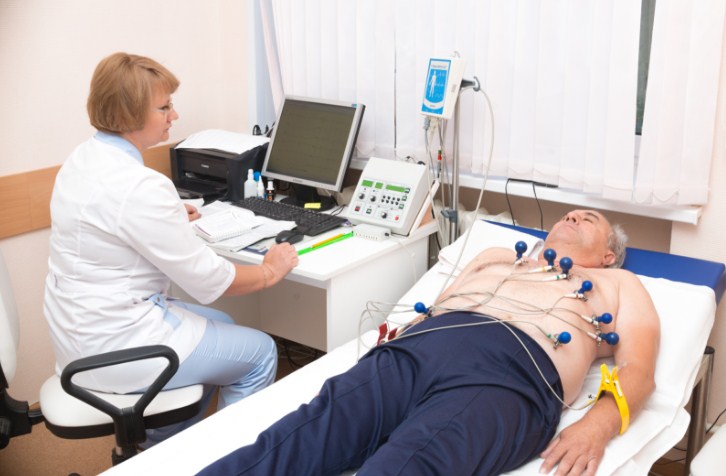
This is an ultrasound examination that is prescribed to accurately assess the patient's condition and identify possible malformations or dysfunctions. Patients are more familiar with another method - an electrocardiogram. Having received a referral for such a procedure, they always ask a lot of questions: how is an echocardiogram performed, what is it, how to properly prepare for the procedure, what is its interpretation.
Description and features of an echocardiogram

It can be used not only for adults, but also for very young children: if it is prescribed by a doctor, you can safely agree and not be afraid of any negative consequences.
This method is based on scanning the patient's chest in the region of the heart using an ultrasound machine.
Such a study allows you to identify even minor deviations from the norm in the work of the most important organ for a person - his heart, to determine its characteristics:
- the size of the heart itself;
- the size of the atria and ventricles;
- the thickness of the atria, septa and ventricular myocardium.
In addition, on ultrasound of the heart, you can set its mass, cardiac output, heart rate and other parameters necessary for an accurate diagnosis.
Such an examination is prescribed by a cardiologist in the following cases:
- when detecting heart murmurs;
- if the patient complains of unstable heart function, sudden interruptions or, conversely, palpitations;
- when an abnormal enlargement of the heart was detected on the radiograph, a change in its shape or localization of the aorta and artery;
- with arterial hypertension, shortness of breath and edema;
- if the patient in the family had cases of heart defects;
- angina pectoris, heart attack, complaints of acute pain in the region of the heart.
An echocardiogram can also be prescribed to pregnant women if they complain of poor health, fainting, dizziness, there are suspicions of developing cardiac pathologies. Diabetes mellitus, rubella during pregnancy, antibiotics, other serious infections suffered by a pregnant woman are also the basis for this study. The transcript will show whether there is really a threat to the mother and child, whether action needs to be taken.
This procedure does not pose any threat to infants. Usually, thanks to modern diagnostic equipment, it is possible to determine deviations from the norm in the fetus even in the womb. After childbirth, an echo is assigned to confirm the diagnosis.
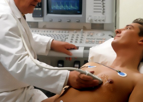
How is an echocardiogram performed?
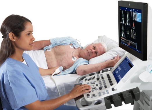
 This procedure has fundamental differences from the electrocardiogram. For its implementation, a special device is used - a converter. The doctor applies it to the patient's bare chest, the device picks up ultrasonic vibrations passing through the heart, and transfers them to a computer, then decoding is performed.
Unlike a traditional electrocardiogram, with an echocardiogram, a doctor can determine:
- whether there are violations of the norm in the work of the heart;
- how well and how much it pumps blood.
The decoding will show whether the patient develops heart failure, and if so, what form and degree it is.
If a patient has an angina attack, a hypertensive crisis or a myocardial infarction, then using this technique, you can determine:
- are there blood clots in the vessels;
- how damaged the valves are;
- whether inflammation of the tissues surrounding the heart has begun;
- whether the heart muscle is enlarged.
Methods for conducting an ultrasound of the heart
Echocardiography can be performed in two ways:
- transthoracic;
- transesophageal.
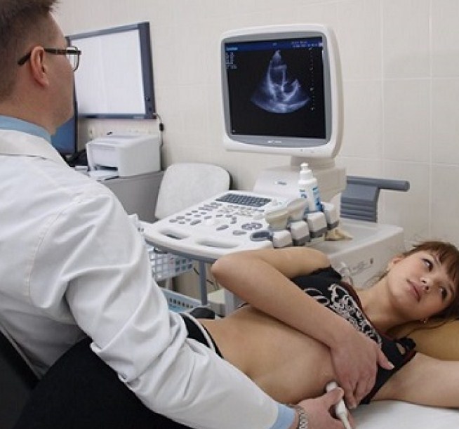
The most commonly used technique is the transthoracic technique. This method has been practiced since the 80s of the last century, therefore for many specialists it is more preferable. Features of this procedure have been described above. The patient is placed in a horizontal position on the couch, after which a sensor is applied to his chest.
The transesophageal technique is more complicated, but gives more accurate results, since the sensor is placed not on the surface of the chest, but in the esophagus.
But in this case, the procedure is much more expensive, it is carried out under anesthesia.
The procedure for transesophageal echocardiography is as follows:
- The patient should not eat or drink before the procedure. If it is scheduled for the morning hours, then the preparation begins the day before. It is necessary to refuse the use of caffeinated drinks and products (chocolate), a light dinner (yogurt or broth) is acceptable. Even water should not be drunk immediately before the examination.
- If the patient must take nitroglycerin-containing drugs, then one day before the echocardiogram, they should be stopped.
- If there are dentures, they must be removed before the procedure.
- For the examination, the patient is placed on the couch, after which he is injected with an anesthetic. General anesthesia is usually used and, in very rare cases, local anesthesia is used if there are contraindications to general anesthesia.
- Then the larynx and pharynx are treated with an anesthetic. Such a measure is needed to protect the mucosa from irritation.
- After that, the patient is connected to equipment that allows you to monitor the work of his heart and lungs, and control the course of the procedure. During general anesthesia, oxygen is also provided.
- A mouthpiece is inserted into the patient's oral cavity, then he turns on his left side and an endoscope with a sensor on the tip is inserted into his throat.
- The endoscope gently advances to the desired depth, after which the doctor examines the heart from different angles for abnormalities.
Obviously, in this way the doctor gets the most complete picture of the state and work of the patient's heart. This is the best option if the patient is obese or the woman has a too voluminous bust, which can make the study difficult. But at the same time, a transesophageal echocardiogram has its own contraindications. These are gastritis, peptic ulcer of the stomach and any neoplasms.
How are echocardiogram results interpreted?
When the study is completed, the ultrasound specialist will print out the photographs and send the patient to the cardiologist with them - and then the results will be deciphered.
The average norm for the parameters is as follows:
- The cavity of the right ventricle at the end of diastole is 1.7 cm, the limit value is 2.6 cm.
- The cavity of the left ventricle at the end of diastole is 4.7 cm, the permissible border movement is 5.7 cm.
- The mouth of the cardiac aorta is 2.7 cm in diameter, the permissible limit value is 3.7 cm.
- The cavity of the left atrium is 2.9 cm, the permissible border movement is 4 cm.
These are the norms of the main indicators that the doctor will determine during the echocardiogram, they are given to guide patients if they have any deviations. But the real decoding is carried out only by a cardiologist. Only he will be able to correctly read the results, explain them and tell the patient how serious his condition is and whether treatment should be started.
Sometimes the indicators of ultrasound of the heart deviate from the norm, but no other signs of cardiac disorders in the examined patient are detected. This most often indicates that the diagnosis was carried out on a low-quality or faulty device. And here it is time to say not only about the great advantages of such a diagnostic method as an echocardiogram, but also about its obvious shortcomings.
What are the advantages and disadvantages of the method
With the help of an echocardiogram, it is really possible to identify almost all cardiac anomalies and detect pathology even at its initial stage of development. A clear image of the chambers and ventricles of the heart is displayed on the screen, the doctor can visually observe the state of the vessels and heart contractions. Today it is one of the most reliable ways to establish the state of the most important human organ.
But at the same time, it also has its drawbacks:
- If the examination is carried out in a regular clinic, then most often the results are unreliable due to the use of old equipment that has already worn out. It is more expedient in this case to go to a private clinic, but also not to any, but to one where good specialists work.
- In such clinics, the cost of an echocardiogram will be several times higher than the cost of a conventional electrocardiogram, which can be done free of charge in a district clinic without any problems. And this is the second drawback of this technique, which often plays a decisive role. Not all patients can afford an echocardiogram, especially repeatedly, if it is required during treatment.
The cost of the examination will depend on such factors:
- clinic level;
- doctor's qualification;
- localization of the clinic - in large cities the price will be higher.