Vertical, horizontal and other types of electrical position of the heart
Content
The human heart constantly generates electrical impulses originating in the sinus node. Further, in the normal state, electrical excitation through the branches and fibers of the conductive nerve bundle (His bundle) moves to the ventricles and the atrium region. This process is expressed by an electric vector with a certain direction. The electrical axis of the heart (EOS) is a projection onto the anterior vertical plane of such a vector. The location of the EOS approximately reflects the location of the heart muscle itself in the chest and is calculated by physicians when undergoing electrocardiography.
Normal indicators of EOS

- normal;
- semi-vertical electrical position;
- vertical;
- semi-horizontal;
- horizontal.
After 40 years, the axis is usually placed at an angle in the range of -30 0 - +90 0 , before this age, the angle is normally formed with indicators from 0 to +105 degrees. In most cases, this indicator is between +30 and +75 degrees.
There are also a number of small deviations that are not pathologies. For example, the diaphragm of asthenic people is located low, so the axis of the heart often deviates to the right, and the heart muscle has a rather vertical arrangement. The heart of overweight people and hypersthenics, on the contrary, is located more horizontally and deviates to the left. For normosthenics, having a proportional ratio of the transverse and longitudinal body size, an intermediate placement of the heart is characteristic.
Things are different with children's indicators of the location of the heart and axis. In newborns, there is a clear shift to the right on the electrocardiogram, and closer to the year the axis usually goes into a vertical position, since the right heart begins to predominate both in weight and in electrical activity. By about 24 months, already in 3 out of 10 children, the normal position is determined.
The establishment of a normal position depends on the rotation of the heart muscle, which reduces the fit of the ventricle to the chest, and the increase in the weight of the left ventricle. In preschoolers and schoolchildren, it is the normal position of the axis that already dominates, the vertical position is less often observed, and the horizontal position is even more rare.
Briefly, the children's EOS norm is expressed as follows:- newborns - +90 - +170 degrees;
- children from one to three years old - the vertical position of the electrical axis of the heart;
- Primary school age and adolescents - the normal position of the EOS in 50% of children.
The data on the angle of placement of the EOS are statistical and approximate. It makes sense to contact specialists only if the data obtained are very different from those described as normal. In any case, for primary confirmation, it is necessary to undergo electrocardiography.
Reasons for EOS deviations
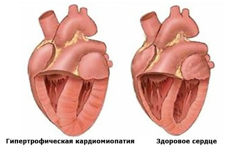
 Deviation of the electrical axis of the heart to the left or to the right is not an independent disease, but sometimes it indicates pathologies that can lead to malfunctions of the heart. So, a shift of the axis to the left occurs in professional athletes, and this is the norm, but often such a deviation occurs due to left ventricular hypertrophy, that is, due to an increase in the weight of the heart and its failures during contractions.
- cardiomyopathy (enlarged myocardium and / or enlarged chambers of the heart) caused by anemia, hormonal failure, coronary disease, cardiosclerosis;
- arterial hypertension with invariably high pressure indicators;
- acquired heart disease that disrupts blood flow inside the heart and increases the load on the left ventricle;
- congenital heart disease, deviating the axis to the left;
- poor conduction in the left bundle branch of His adversely affects the contraction of the left ventricle;
- atrial fibrillation not only deviates the axis, but also develops a non-sinus rhythm.
- diseases of the respiratory system, for example, chronic inflammatory disease of the respiratory tract, bronchitis with obstruction, emphysema;
- heart disease affecting the tricuspid valve and pulmonic valve.
EOS diagnostics
 To identify the causes of the deviation after the detection of axis displacement, the cardiologist or the attending doctor prescribes additional examinations:
- Ultrasound examination of the heart muscle, which evaluates anatomical changes in the most detailed way and determines ventricular hypertrophy. This method is important to use in the diagnosis of newborns to identify cardiac pathologies of a congenital nature.
- Electrocardiography with stress (treadmill test and bicycle ergometry) to determine ischemic myocardial disorders, which are one of the possible causes of EOS displacements.
- Holter monitoring of the ECG is prescribed if other studies have shown both an axis shift and a non-sinus rhythm, which is pathological.
- Radiography is indicated if myocardial hypertrophy with enlarged cardiac shadow is present.
- Coronary angiography is performed to determine the exact nature of coronary artery lesions in coronary artery disease.
Treatment, prognosis and potential consequences
As noted earlier, the very fact that the EOS has a deviation in one of the parties is not directly a disease, and therefore does not require specific special treatment. The displacement of the EOS is rather a criterion, potentially suggesting the presence of one of several possible pathologies of the heart.
If such a pathology is eventually detected due to the fact that an axis deviation was detected on the ECG, it is necessary to go through the standard procedural steps:- Contact the attending therapist, providing all the results of the examination already passed.
- Get a referral to a cardiologist. Also provide at the appointment a medical history, an electrocardiogram and other results of the tests passed, if necessary.
- Sign up and go through all the studies recommended by the attending physician, consult with specialists from other fields of medicine, if the diagnosis requires it. Strictly follow the treatment plan and medication plan chosen by your doctor.
Separately, it is worth considering how the axis shift affects children.
 From birth to 3 months, the axis is deviated to the right due to the fact that the right ventricle is larger in both size and degree of activity, but after three months of age and the axis changes to a right angle, the following changes can occur:
- rotation of the heart muscle;
- reduction in the area of contact of the right ventricle with the chest;
- an increase in the mass of the heart in the left section;
- the rightogram goes to the normogram.
After two years of age, the normal position of the EOS should, as a rule, form. However, even if there is still a shift to the right side, vertical, horizontal or intermediate position, a specific diagnosis cannot be made without additional examinations.
In this case, it is most likely that the displaced EOS is simply related to the anatomical features of a particular person.
But when you really should be on your guard, it is when detecting a rightogram in patients with lung diseases, or a leftogram in people with hypertension. In these diseases, the displacement of the axis of the heart may indicate the progression of the primary disease.
If the main diagnosis has not yet been made, but the ECG has already revealed a strong deviation of the EOS and cardiac symptoms, then the patient is definitely recommended to undergo a full examination.